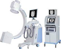
Features
- With a compact appearance, and easy to operate.
- Unique base electric auxiliary support arm design, it’s more security for using.
- A unique hand-held controller design, convenient to operate.
- With a high-quality knockdown X-ray generator to reduce radiation.
- With the Perspective KV, MA automatically track fluoroscopy to make the image brightness and clearness optimum.
- Toshiba image intensifier three view, the quality is stable and reliable, a good image clarity.
- Image acquisition and processing workstation
Registration: registration, medical records, Worklist
Collection: Start collection; prepare video, reset, horizontal mirror, vertical mirror, window adjustment, magnifying glass, negative image
Open silhouette, edge enhancement, recursive noise reduction
Processing; four windows, nine windows, sharpening, horizontal mirror, vertical mirror, text annotation, length measurement report: save, preview, expert template
Dicom features: Dicom browsing, web service
Automatically track fluoroscopy to make the image brightness and clearness optimum; Installation of dense grain grids, to further enhance image sharpness
- Automatic dynamic motion detection when fluoroscopy to avoid movement-related blurring, no silhouette
III. Configuration
1, New (with electric auxiliary support arm) C-arm host 1 set
2, High-frequency high-voltage X-ray generator and high-frequency inverter power supply(5KW、40KHZ、120KV) 1 set
3, Toshiba 9-inch image intensifier three view(9inch/ 6 inch / 4.5 inch)1 set
4, Imports of super-low-light digital camera 1024×1024 1 set
5, CCU in the high-definition progressive output control system 1 set
6, Dense grain grids 1 set
7, Electric adjustable beam collimator 1 set
8, 19 inch LCD display 2 sets
9, hand-held controller 2 sets
10, Foot switch for exposure 2 sets
11, Red-cross positioned 1 set
10, Digital work station with printer 1 set
- Technical Specifications
| PARAMETER | SPECIFICATION | |
| Fluoroscopic
Capacity |
Photography Max rated capacity | 5KW |
| Fluoroscopic ax rated capacity | Tube Current 4mA, Tube Voltage 120kV | |
| Automatic Fluoroscopy | Tube Voltage: 40kV~120kV adjust automatically
Tube Current: 0.3mA~4mA adjust automatically |
|
| Manual Fluoroscopy | Tube Voltage: 40kV~120kV Continuous
Tube Current: 0.3mA~4mA Continuous |
|
| Pulse Fluoroscopy | Tube Voltage:40kV~120kV Continuous
Tube Current: 0.3mA~8mA Continuous (1,) intelligent frequency conversion technology is more advanced (2,) improves the quality of single-frame images and reduces the amount of radiation (3,) the tube is protected, continuous working time is extended (4,) frequency: 0.5 ~ 8pps (frame / s) pulse length |
|
| Photography Tube Voltage and mA | 40 kV~120 kV 20-100mA 1.0mAs~180mAs | |
| Plate holder Size | 200mm×250mm(8″×10″) or
250mm×300mm(10″×12″) |
|
| X-ray Tube | X-ray Tube Special for High Frequency | Fixed anode Dual-focus, 0.3/1.5,
Inverter Frequency: 40KHz |
| Anode capacity: 35KJ (47KHU)
Tube thermal capacity: 650kJ(867kHu) |
||
| Video
System |
Image Intensifier | Image Intensifier made by TOSHIBA (9″)
Three view(9inch/ 6 inch / 4.5 inch) E5764SD-P3 Image definition indicators 12 bit |
| CCD Video camera | 1 Mega Ultra low-light CCD camera | |
| Monitor | 19”LCD monitor *2:
Resolution 1280*1024, |
|
| CCU(central control) | Recursive Filter: K=8, 8 images storage, image upright, image overturn, positive & negative image;LIH(last image freeze, and OSD (monitor display) | |
| Structural performance | Directive wheel | ±90°revolution, can freely change the moving direction of the unit. |
| C-arm | Forward and Backward Movement: 200mm
Up and down:400mm Revolution around Horizontal Axis: ±180° Revolution around Vertical Axis: ±15° Focus screen distance: 960mm C-arm opening: 740mm C-arm arm depth: 640mm Slide along the orbit: 120°(+90°~ -30°) |


